MRI can leak mercury from dental fillings
Research published in the pages of the journal "Radiology" showed that levels of poisonous metal were much higher after exposure to strong pol magnetic, whichore can generate the latest devices of this type, approved for use last year.
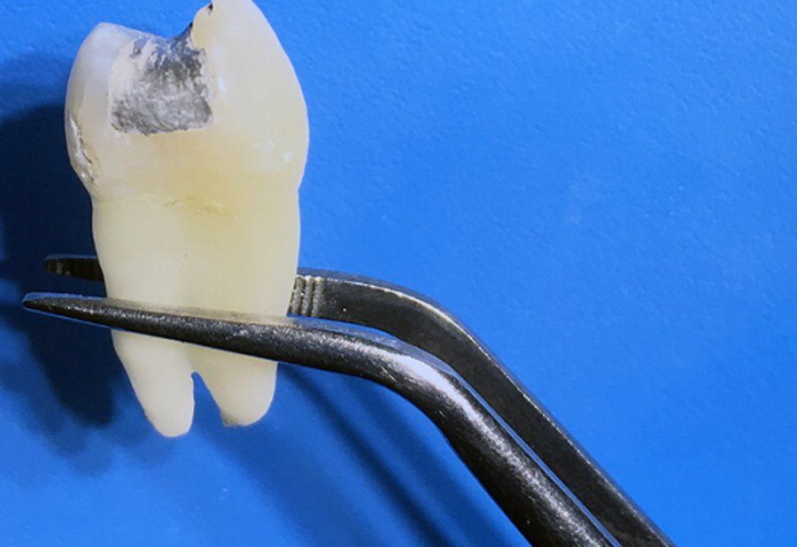
Amalgam is ogolna name of alloysoin metals, where the primary component is mercury, which is toxic to humans. In dental amalgams, the liquid metal is more than 50 percent. Although one can find roAlso silver, tin, copper, cadmium and sometimes zinc. For many years it was used, as a cavity fillingoin dentition. In today’s dentistry, these types of fillings are no longer used, precisely because of the risk of exposure to doctors and patientsoIn contact with toxic metal. However, there are still millions of osob all over the world have amalgam fillings in their teeth. And this can be quite a problem for them.
Although mercury is highly toxic to humans, amalgam fillings are considered safe. The technique of inserting them excludes any penetration of. Of course, if carried out properly. The cured amalgam binds to the chemical structure of the tooth after 48 hours. However, the strong magnetic field may cause leakage.
MRI machines are increasingly available in hospitals to provide detailedondings for imaging complex structures, such as mozg or in diagnosing strokeow or cancerow. Most such devices are between 1.5 and 3 Tesla, but modern MRI machines can be as powerful as 7 Tesla.
To test the new machines, Dr. Selmi Yilmaz, a dentist at Akdeniz University in Turkey, along with a team of cooemployeeow, created artificial cavities in removed teeth and filled them with amalgam. After the fillings were cured, the teeth were placed in an artificial saliva solution and then exposed for 20 minutes to a 1.5 T magnetic resonance camera and 7 T. Tooth control groupow with an amalgam filling was not exposed to a magnetic field, but placed in artificial saliva.
Previous studies have already shown that mercury leakage from amalgam fillings can occur during MRI examinations. But when new devices with much greater power appeared on the market, thanks to which theorym imaging is more detailedołowe, niepokoj wzrosł. The author of the paper admitted that the new apparatuses were met with ogolion’s share of satisfaction because they can visualize previously unseen anatomical details, but no one has studied them for use on patients with old fillings.
The experiments showed that the mercury content in the artificial saliva solution after the magnetic resonance force of 7 T was almost four times higher than in the case of a scanner with lower power. It amounted to 0.67 ppm (parts per million) in porown comparison with 0.17 ppm for probki exposed to a magnetic field of 1.5 T and 0.14 in the control group.
– Although it is unclear how much of the released mercury is absorbed by the body, the findings indicate that amalgam fillings may pose a risk not only to the patientow, but roAlso for the personnel operating the apparatus – Yilmaz said, adding that the 1.5T MRI scan is safe and patients with old amalgam fillings can undergo the scan without fear.


